Created by Itzhak Brook MD a physician and a laryngectomee. It contains information about head and neck cancer, life after laryngectomy, and manuscripts and videos about Dr. Brook's personal experiences as a patient with throat cancer. It has information about side effects of radiation and chemotherapy; methods of speaking; airway, mucus, stoma, voice prosthesis; eating and swallowing; medical, dental and psychological issues; respiration; anesthesia; travelling; and COVID-19.
"My Voice"
Order a paperback or Kindle Edition or e-book of "My Voice: A Physician's Personal Experience with Throat Cancer," the complete 282 page story of Dr. Brook's diagnosis, treatment, and recovery from throat cancer.
Order a paperback or Kindle Edition or e-book of "The Laryngectomee Guide," the 170 page practical guide for laryngectomees.
To obtain suggestions for laryngectomees how to cope with COVID-19 pandemic click the Laryngectomee Newsletter link.
Side effects of radiation treatment in head & neck cancer
Radiation therapy (RT) is often used to treat head
and neck cancer.It can be used as the only treatment, in combination with chemotherapy (chemoradiation therapy), or after surgery (adjuvant radiation therapy). The goal of radiation therapy is to kill cancer cells. Because these cells divide and grow at a faster rate than normal cells they are more likely to be destroyed by radiation. In contrast, although they may be damaged, healthy cells generally recover. Unfortunately radiation treatment causes short and long term side effects. RD can damage blood vessels that nourish muscles, nerves, and bones that can result in a progressive condition called "radiation fibrosis syndrome", which causes a variety of complications affecting nerve, muscles, and bones. Some side effects (e.g., nausea, mucositis) are generally more pronounced in those who receive radiation in combination with chemotherapy.
Radiation Fibrosis Syndrome video from Sloan-Kettering Cancer Center
RT can be administered in several ways:
Organ
preservation - radiation
is aimed at the tumor site (with or without chemotherapy) is used in an attempt
to cure the disease without surgically removing the larynx. However, this is not
always an option because of the size and location of the tumor and the
recommendation is to proceed directly to surgery.
Palliative
treatment - radiation
(with or without chemotherapy) is given in an attempt to prolong life when the
tumor is too large and/or inoperable and cure is highly unlikely.
Radiation
after surgery - radiation is given after surgery to destroy any local residual cancer cells
that may spread to other organs such as the lung, liver, or brain.
Reirradiation for recurrent cancer -radiation is administered for recurrence of head and neck cancer in a previously irradiated area. Repeat irradiation with systemic therapy is a potentially curative
option. Long-term disease-free survival has been observed, albeit with the risk
of significant, possibly life threatening, late complications.
Types
of radiation therapy
Most patients with for head and neck cancers are treated withexternal beam radiation therapy(using X-rays or gamma rays). The current standard of care is
to use intensity-modified RT (IMRT). This method adjusts the beams to maximize radiation to cancerous tissue and not to normal
tissue. This reduces side effects of RT. An individual face
mask is made for each patient to insure accurate delivery of radiation. The
number of treatments a person may get depends on the cancer type. Some patients
get radiation only a single time while others get radiation once a day, 5
days a week, for up to 7 weeks.
Other
methods of radiation include:
Brachytherapy - implanting radioactive source close to the cancer
TomoTherapy - combines precise 3-D imaging from computed tomography (CT) scanning with highly targeted radiation
beams delivered precisely to the cancer while minimizing surrounding tissues
damage)
Conformal radiation therapy - radiation beams are shaped to match the tumor’s 3 dimensional
picture based on CT and/or magnetic resonance imaging (MRI) scans)
If RT is recommended the radiation oncologist sets up a treatment plan
that includes the total dose of radiation to be administered, the number of treatments to be given, and their schedule. These are based on the type and location of the tumor, the patient's general health, and other present or past treatments. For early stage disease, doses of 66-74 Gy are generally administered.
The likelihood and severity of complications depends on a number of
factors, including the total dose of radiation delivered, over what time it was
delivered and what parts of the head and neck received radiation. The side effects of RT for head and neck cancer are divided into early (acute) and long term (chronic) effects. Early side effects occur during the course of therapy and during the immediate post therapy period (approximately 2-3 weeks after the completion of a course of RT). Late effects can manifest any time thereafter, from weeks to years later.
I described my own experiences getting RT in my book “My Voice” in chapter 4 ”Getting irradiated”, and chapter 5 "Life after irradiation". A lecture about life challenges after laryngectomy that includes discussion of late side effects of radiation can be viewed on YouTube.
Patients are
usually most bothered by the early effects of RT, although these will generally
resolve over time. However, because long term effects may require lifelong care
it is important to recognize these in order to prevent them and/or deal with
their consequences. Knowledge of the radiation side effects can allow their
early detection and proper management. Individuals with head and neck cancer should receive counselingabout the importance of smoking cessation. In addition to the fact that smoking is a major risk factor for head and neck cancer, the risk of cancer in smokers is further enhanced by alcohol consumption.Smoking can also influence cancer prognosis. When smoking is continued both during and after RT, it can increase the severity and duration of mucosal reactions, worsen the dry mouth (xerostomia), and compromise patient outcome. Patients who continue to smoke while receiving RT have a lower long-term survival rate than those who do not smoke.
Emotional needs during Radiation therapy
Up to half of patients receiving
radiation therapy may experience anxiety and distress especially during the
first visits to radiation oncology. Radiation therapy provokes high anxiety,
with patients reporting fear of radiation and that being in an oncology
department reminds them of their life‐threatening condition.
Radiation therapists (RTH) are the main
health professionals that are in direct daily contact with patients during radiation
treatment, placing them in a unique position to explore and deal with patients’
psychosocial needs. Their role incorporates patient education, including
explanation and co‐ordination of procedures and
appointments, and providing advice regarding personal care during treatment. In
fulfilling these roles, RTHs need to spend time with patients to ensure their
information needs are met and that they are willing to proceed with treatment. Consequently,
RTHs have a role in providing psychosocial support to patients. RTH–patient
interactions can reduce patient anxiety through effective communication,
forming relationships, acknowledging patients as individuals and provision of
education/information.
RTHs prepare patients for the procedure
through education and information before the start of treatment. Adequate
preparation can reduce patient anxiety as well as recovery time and
complication rates. Furthermore, RTHs interact with patients daily, and
throughout treatment are able to tailor information to suit individual
patient's changing needs and to involve patient's in their own care. The RTH–patient
communication also enables RTHs to consider whether to involve families and caretakers
in education/information sessions which may improve the overall patient experience
and potentially reduce patient and family anxiety.
It is advisable that patients and family
members utilize the guidance and assistance provided to them by the RTH. This can
reduce anxiety and guide the patient through the radiation therapy and afterwards
as they deal with the treatment’s short and long term side effects.
Early side effects
Early side effectsinclude inflammation of the oropharyngeal mucosa (mucositis), painful swallowing (odynophagia), difficulty swallowing (dysphagia), hoarseness, lack of saliva (xerostomia), increased mucus production, orofacial pain, Laryngeal radionecrosis, dermatitis, hair loss, nausea, vomiting, inadequate nutrition and hydration, and weight loss. These complications can interfere with, and delay treatment. To some degree these side effects occur in most patients and generally dissipate over time.
The severity of these side effects is influenced by the amount and method by which the RT is given, the tumor’s location and spread, and the patient’s general health and habits (i.e. continued smoking, alcohol consumption).
Skin damage (radiation-induced dermatitis)
RT can cause a sunburn-like damage (radiation dermatitis) to the skin which can be further aggravated by chemotherapy. It is one of the most common side effects of RT and can cause pain and discomfort. The dermatitis depend upon the radiation dose and can be mild, moderate and severe. The severity of dermatitis and healing time are significantly increased in patients taking radiosensitizing agents.
It is advisable to keep the irradiated area clean and dry, wear loose-fitting clothes to avoid friction injuries,wash the skin with lukewarm water
and mild soap (preferably synthetic soaps), and avoid exposure to potential chemical irritants, skin irritants such as perfumes and
alcohol-based lotions, direct sun and wind, and local application of lotions or ointments prior to RT that might change the depth of radiation penetration.
There are a number of skin care products that can be used during radiation treatment to lubricate and protect the skin. These include aloe vera-based gels and water-based lotions. Although such preparations may provide symptom relief, none promotes or accelerates healing of the radiation-induced dermatitis. Topical
corticosteroids can be used during and a several weeks after completion of
treatment may prevent severe radiation dermatitis and reduce discomfort and
itching.
The management
of radiation dermatitis is guided by the severity of skin damage and includes
general skin care measures, prevention and treatment of secondary skin
infection, and the use of dressings.
Mild dermatitis starts improving with 10 days after completing of radiation, while severe dermatitis is associated with prolonged inflammation and healing time, resulting in skin fibrosis.
Skin cancer can rarely develop at the irradiated areas.
Wearing adhesive heat and moisture exchanger (HME) housing is not recommended during RT and the recovery period as the skin around the
stoma usually become inflamed.
Hair
follicles are very sensitive to radiation, and the treatments can cause hair
loss. Most individuals observe hair loss within the treatment area about three
weeks after the beginning RT. Hair loss may be temporary or permanent,
depending on the total amount of radiation received and other treatments such
as chemotherapy. When hair loss is temporary, it will likely re-grow within 3
to 6 months after treatment is complete. The re-growth of hair is often thinner
or of a different texture.
Some
individuals elect to have their hair cut short prior to starting RT. Those who
wish to wear a wig, are advised to select it prior to losing their hair in
order to match color and style.
The
scalp is sensitive to radiation, especially following hair loss. The skin may
become pink, tender or inflamed - like a sunburn. Following 2-3 weeks of
treatment, the scalp may become dry and itchy. Appropriate special cream can be
prescribed and applied to these areas.
The
dry, irritated scalp is a temporary condition and start improving about two
weeks after RT is complete. When indicated, medications can be administered to
relieve discomfort and itching.
The scalp
reaction can be minimized during the treatment by:
Avoiding
frequent shampooing and using a mild shampoo (such as baby shampoo) without any
perfumes.
Washing
the scalp with warm water only and avoiding rubbing and scratching.
Drying
the washed area by patting with a dry soft towel.
Avoiding
excessively combing or brushing the hair.
Avoiding
the use of hair spray, oils or creams.
Avoiding
the use of heat sources (including hair dryers, rollers or curling irons).
Avoiding
perming or coloring the hair until about 4 weeks after RT is complete.
Protecting
the head from the sun, cold and wind by wearing a head covering (i.e., cap, scarf, and cotton hat).
Losing one's hair can be upsetting. Wearing a wig, scarves, turbans, bandanas, and hats having a short hair cut can be helpful.
Hair loss
Dry mouth (xerostomia)
Saliva is produced by three pairs of salivary
glands: The parotid (produces about 30% of the saliva), sublingular (<5%),
submandibular (60%). There are also approximately 600 to 1000 minor salivary glands. The salivary glands are directly effected by the radiation treatment resulting in radiation-induced sialadenitis. The loss of saliva production (or xerostomia) is the most common long-term complication of radiation therapy, and is related to the administered irradiation dose and the volume of salivary tissue irradiated. In most individuals, the saliva becomes more viscous,
thick and stringy, and difficult to expectorate; its buffering capacity is
reduced, and its pH shifts from neutral to acidic which increases in dental
decay bacteria and initiates dental demineralization process. Xerostomia generally stars 4 weeks after
initiation of RT and generally takes up to eighteen months to improve.
Prevention
of permanent salivary gland damage can be attempted in selected patients by
using parotid-sparing
intensity-modulated radiation therapy (IMRT), reduce
the radiation dose to the submandibular and minor salivary glands (if oncologically feasible),submandibular salivary gland surgical transfer, and administration of
amifostine (a radiation protective organic thiophosphate medication).
Although xerostomia generally improves with
time, it is often a permanent problem that can adversely impacts quality of
life.
These measures can help in coping with xerostomia:
Drinking adequate fluids, frequent
sipping or spraying of the mouth with water; sucking on ice chips and /or
sugar-free popsicles; using sugarless gum and sugarless hard candy can help
stimulate saliva, and rinsing and gargling with diet ginger ale or a weak
solution of salt and baking soda are helpful to refresh the mouth, loosen thick
oral secretions, and alleviate mild pain.
The use of saliva substitutes, or
stimulation of saliva production from intact salivary glandular tissues by
taste/mastication, pharmacological sialogogues (a drug that increases the flow rate of saliva), acupuncture, avoiding smoking and all products that contain caffeine or
alcohol, using a bedside humidifier at night, and raising the head of the bed
can be helpful.
Soft and moistened foods, thick soups, mashed potatoes, puddings, and milkshakes are easier to eat and swallow. For more information see Xerostomia at the Late side effects section below.
Acceleration of periodontal disease
Patients
who experience low function of their salivary gland and xerostomia must
maintain excellent oral hygiene to minimize the risk of oral lesions.
Periodontal
disease can be accelerated and caries can become rampant unless preventive
measures are instituted. Multiple preventive strategies should be considered. This
evolves performing systematic oral hygiene at least 4 times per day (after
meals and at bedtime) which includes:
Brushing
teeth (if soreness of oral mucosa and trismus are present, a small ultra-soft
toothbrush can be used).
Using
a fluoridated toothpaste when brushing
If
toothpaste makes one's mouth sore, brush with a solution of 1 teaspoon of salt
mixed with 4 cups of water.
Flossing
once daily.
Applying
a prescription-strength fluoride gel at bedtime to prevent caries.
Rinsing
with a solution of salt and baking soda 4 to 6 times a day (½ tsp salt and ½
tsp baking soda in a cup of warm water) to clean and lubricate the oral tissues and
to buffer the oral environment.
Sipping
water frequently to rinse the mouth and alleviate mouth dryness.
Use
of topical fluoride has demonstrable benefit in minimizing caries formation.
During radiation treatment, it has been recommended that mouth guards be filled
with topical 1% sodium fluoride gel and placed over the upper and lower teeth.
The appliances should remain in place for 5 minutes, after which the patient
should not eat or drink for 30 minutes.
Mouth guards can be filled with topical fluoride gel
Alterations in taste (dysgeusia)
Radiation can induce changes in taste as well as tongue pain. Foods
can alternately taste too bland or too spicy due to the tongue's limited taste
receptors.Some foods may taste different than they did in the
past, some foods may taste bland, or every food may taste the same.
Specifically, bitter, sweet, sour, and salty foods may taste different, and some
people may have a metallic or chemical taste in their mouth, especially after
eating meat or other high-protein foods.
The sense of taste may also be affected by impaired smelling. These side effects can cause food aversion (dislike), further decrease food intake, and contribute to weight loss.
RT
as well as chemotherapy can impair the
sense of taste because of their effects on the taste buds in the tongue and nasal
epithelium receptors. Additional factors that may contribute to an altered
sense of taste include a bitter taste from chemotherapy drugs, poor oral
hygiene, infection, and mucositis.
Taste
changes and tongue pain caused by RT usually begin to improve three weeks to
two months after the end of treatment. Improvement may continue for about a
year, but the sense of taste may not entirely return to the way it was before treatment,
especially if there is damage to the salivary glands.However, alteration in food choices and
preparation may minimize the effect of change of taste on the ability to
consume adequate and nutritious foods.
In
most instances, there are no specific treatments for taste problems.
These
tips may help to cope with taste changes:
Choosing
foods that smell and taste good, even if the food is not familiar.
Eliminating
cooking smells by using an exhaust fan, cooking on an outdoor grill, or buying
precooked foods. Cold or room-temperature foods also smell less.
Eating
cold or frozen food (i.e., frozen yogurt, ice cream), which may taste better than hot foods.
Using
plastic utensils and glass cookware to lessen a metallic taste.
Trying
sugar-free, mint gum or hard candies (with flavors such as mint, lemon, or
orange) to mask a bitter or metallic taste in the mouth.
Trying
other protein sources (such as poultry, eggs, fish, peanut butter, beans, or
dairy products) if red meats don't taste good.
Marinating
meats in fruit juices, sweet wines, salad dressings, or other sauces.
Flavoring
foods with herbs, spices, sugar, lemon, or sauces.
Not
eating one to two hours before and up to three hours after chemotherapy to
prevent food aversions caused by nausea and vomiting. Additionally, avoiding
favorite foods before chemotherapy helps prevent aversions to those foods.
Rinsing
with a salt and baking soda solution (½ teaspoon of salt and ½ teaspoon of
baking soda in 1 cup of warm water) before meals, which may help neutralize bad
tastes in the mouth.
Keeping
a clean and healthy mouth by brushing frequently and flossing daily.
Considering
zinc sulfate supplements, which may help improve taste in some people. However,
one should consult with their physician before taking any dietary supplements,
especially during active treatment.
Photobiomodulation therapy employing light at
red and near-infrared wavelengths has been used for the treatment of taste alteration.
Inflammation of the oropharyngeal mucosa (mucositis and odynophagia)
Radiation, as well as chemotherapy, damage the oropharyngeal mucosa resulting in mucositis, and odynophagia (pain with swallowing) which develops gradually, usually 2-3 weeks after starting RT and gets better starting 5 weeks after RT has
ended.Its incidence and severity depend upon the field, total dose and duration of RT. Chemotherapy can aggravate the condition. Factors the
increase the risk of mucositis include: poor oral hygiene; poor dental health;
smoking or using tobacco products, including chewing tobacco; using alcohol;
not drinking enough fluids; dry mouth; female gender; younger age; and having
suffering from another disease (i.e., diabetes, HIV/AIDS, kidney disease).
The World
Health Organization (WHO) scale of mucositis combines both subjective and
objective measures of oral mucositis:
Grade 0 = No
oral mucositis
Grade 1 =
Erythema and soreness
Grade 2 =
Ulcers, able to eat solids
Grade 3 =
Ulcers, requires a liquid diet (due to mucositis)
Grade 4 =
Ulcers, alimentation not possible (due to mucositis)
The mouth is
the most common site for mucositis, but it can also occur in the throat. The
parts of the mouth that are most likely to be affected are the inside of the
cheeks and lips, and the tongue (especially the sides and bottom). Oral
mucositis can range from mild to severe. In mild cases, the sores may be small
and only cause a little discomfort. In the most severe cases, sores are large,
widespread, and extremely painful.
Symptoms include: mouth, gums, tongue, and throat are sore and may be
covered by a white or yellow buildup;
pain or discomfort; red, shiny, or swollen areas in the mouth; mouth
bleeding; white patches or pus in the mouth; difficulties and pain when swallowing saliva, food and drinking; difficulties in speaking; and increased
mucus or thick saliva.
Painful mucositis can interfere with food
intake and nutrition, as well as swallowing saliva. Some individuals need to spit
their saliva rather than swallowing it.
Management includes meticulous oral hygiene, dietary modification, and ingestion of topical anesthetics combined with an antacid and antifungal suspension ("cocktail") before eating. Spicy, acidic, sharp, or hot food should be avoided, as well as alcohol. Reducing the pain on swallowing can ease and increase food and liquid consumption. Although mucositis is not an infection, secondary bacterial, viral (i.e., Herpes), and fungal (i.e., Candida or thrush) infections are possible. Control of the pain (using opiates or gabapentin)may be needed.
Currently,
various strategies and agents are available for the prevention of
mucositis, including routine oral care, mucosal surface protectants,
anti-inflammatory drugs, growth factors, certain antimicrobial formulations,
laser therapy, oral cryotherapy, and specific natural and miscellaneous agents.
These approaches encompass a diversity of mechanisms, but the results have been
controversial, and the optimal prophylaxis remains unknown.
Mucositis can lead to nutritional deficiency. Those who experience significant weight loss or recurrent episodes of dehydration may require feeding through a gastrostomy feeding tube.
When
getting radiation therapy to the head or neck, one needs to take good care of their
teeth, gums,
mouth, and throat.
Although mucositis
is not an infection, open mouth sores, reduce saliva production, weakened
immune system, poor diet and poor oral hygiene raises s the risk of local and
systemic bacterial, viral, and fungal infection. Individuals with neutropenia
are at increased risk of sepsis. Severe mucositis can disrupt the treatment and
reduce the quality of life.
Before You
Begin Treatment
While most head
and neck cancer patients will experience some degree of mucositis during
treatment, there are some things can do before beginning treatment to reduce
the chance of developing the condition.
Getting examined and treated by a dentist who is
knowledgeable and experienced working with cancer patients.
Correcting
any dental issues prior to starting to treatment. Idealy any dental work,
including fillings or extractions, should be done 4-6 weeks before starting
radiation or chemotherapy.
Dental
or another oral prosthetic, should fit properly.
Asking
one’s medical team about medications or treatments that may reduce the risk of
developing oral mucositis.
These
may include:
•Photobiomodulation therapy – a type of low-level laser therapy that may prevent oral mucositis if used before
you begin cancer treatment. This therapy is new to for treatment of
mucositis, and shows great promise without side effects
•Amifostine
(Ethyol®) – a drug that protects the salivary glands and oral mucosa against
damage caused by radiation.
•Benzydamine– a topical nonsteroidal anti-inflammatory agent).
•Asking
one’s doctor about having a feeding tube placed before treatment starts. That
way, it is already in place when eating becomes too difficult. Waiting to get a
feeding tube until it is needed can result in a medical and nutritional crisis,
but it is a crisis that can be prevented by planning ahead. The tube is easily
removed once it is no longer needed.
Management
includes meticulous oral hygiene, dietary modification, and ingestion of
topical anesthetics combined with an antacid and antifungal suspension
("cocktail") before eating. Spicy, acidic, sharp, or hot food, and
alcohol should be avoided. Reducing the pain on swallowing can ease and
increase food and liquid consumption. Secondary bacterial, viral (i.e.,
Herpes), and fungal (i.e., Candida or thrush) infections are possible. Control
of the pain (using opiates or gabapentin) may be needed. (See below)
Once oral
mucositis begins, there are steps you can take to help relieve pain and
symptoms. Some basic tips include:
Stop smoking and/or chewing tobacco
Avoid alcohol
Drink a lot of fluids
Make sure your diet includes lots of protein, which helps your body
rebuild and repair itself
Eating and
Drinking
Although it may
be painful, it is important to make sure you’re eating and drinking enough; eating smaller, more frequent meals throughout
the day. To
reduce pain and discomfort while eating, choose foods that soothe the mouth,
such as:
Cold foods, including popsicles, frozen fruit, and ice cream
Soft, mild foods such as cottage cheese, smoothies, and yogurt
Well-cooked, soft meals such as potatoes, macaroni and cheese,
casseroles, stews, and pasta in white sauce
Drink through a straw to avoid sore spots
Eat sugar-free candy or chew gum to help keep your mouth moist.
Moisten food with gravies and sauces to make it easier to eat.
Stay away from
foods that could irritate your mouth including:
Acidic foods, such as citrus fruit, tomatoes, peppers, and vinegar
Spicy foods
Crunchy or hard foods such as crusty bread, pretzels, and chips
Hot foods
Alcohol and carbonated drinks
Sugary snack
Oral Care
It is important
to take good daily care of your mouth, especially during treatment and if
mucositis develops.
Brush
your teeth. Tooth brushing helps keep the mouth moisturized, and it helps
prevent infection.
Use
a soft-bristle toothbrush. Some patients find it easier and more comfortable to brush with a foam brush with an antibacterial rinse. Do not use lemon or
glycerin swabs.
Brush
gently at least twice daily. Some patients find it helpful to brush every 4
hours and at bedtime.
Use
a gentle, mild-tasting toothpaste with prescription-strength fluoride, such as
Prevident. Do not use whitening toothpastes. If flavored toothpastes irritate the mouth, brushing with plain baking soda ora solution of 1 teaspoon of salt mixed with 4 cups of water is less irritating.
Floss
gently once a day.
Rinse
your mouth frequently with a product such as Peridex or Periogard to prevent
infection.
Salt
water also provides many of the same benefits of moisturizing and cleaning.
Be
sure to avoid mouthwashes that contain alcohol that can dry and irritate mouth tissues.
Avoid
using toothpicks which can cut your mouth.
Use
lip balm or moisturizer to keep your lips moist.
Avoid
using Vaseline because it can promote bacteria growth.
Rinse
your mouth before and after meals and at bedtime with a baking soda rinse to
relieve pain and clean out mucus buildup. A good recipe is: ½ tsp salt, 1 tsp baking soda in 1 quart of wate. Be sure to RINSE and SPIT – do not swallow! Prepare a fresh batch each day. Additional
tip: If the salt makes the rinse too painful, reduce the amount until it is no longer
uncomfortable.
If the gums are sore, dentures should be worn only
while eating. Dentures should not be worn If sores are severe. Loose or fit
poorly dentures should not be worn. Poorly fitting dentures may rub and irritate
the mouth leading to more sores. Remove and
clean the dentures every night or as directed by the dentist.
Pain Control
·Pain
from mucositis can range from mild to severe. To help manage pain, try the
following:
Suck
on ice chips or popsicles and sip cool drinks often throughout the day.
Clean
your teeth and mouth after meals and before applying topical coating agents or
medication for mouth sores.
Topical
pain relievers, including lidocaine, benzocaine, or dyclonine hydrochloride may
provide temporary relief.
In
severe cases, you may need a prescription for a pain medication ( i.e., opiates, gabapentin)
Many
resources suggest using “magic mouthwash” – a
mixture of lidocaine (a pain reliever), diphenhydramine (an
antihistamine and anti-inflammatory), and Maalox. “Magic mouthwash” should be
used with caution because Maalox can cause drying of the lining of the mouth,
which can make mucositis worse.
Topical medications to manage mucositis, including
corticosteroids (anti-inflammatory drugs) mouthwas, and anti fungal agents.Other medications that may be prescribed are mucoadhesive
hydrogel, Palifermin, Gelclair® and Zilactin®. These medications coat and protect the
mucus membranes and nerves exposed by sores. The medical team may also
recommend photobiomodulation therapy, a type of light therapy that may reduce
the severity or duration of oral mucositis.
Viscid,
copious mucus production is a major problem for many patients with severe
mucositis. The mucus causes queasiness and gagging and contributes to difficulty
in maintaining adequate hydration and nutrition.
The
secretions can be managed by:
Regular mouth rinsing with salt and soda solution, and taking oral guaifenesin in the early-phase of mucositis.
Later-phase
thickened or larger-volume mucus may respond to combination narcotics and
anticholinergic drying agents present in some cough preparations.
Treatment
with mucus drying medications include: an antihistamine, and scopolamine transdermal
patch.
Lysine
supplements may be helpful.
Elevation
of the head of the patient’s bed 30° can reduce edema and protect the airway.
A
cool mist vaporizer may help lubrication and expectoration.
Lorazepam
can help prevent repeated gagging and nausea.
Suction
machine can be useful, especially after surgery when effective gargle is
difficult.
The
duration of mucositis is proportional to the degree of mucosal stem cell
depletion. Radiation-induced mucositis may take weeks to months to heal depending on mucosal stem cell
recovery. Excessive depletion may prevent healing and lead to a chronic open wound
recognized as “soft-tissue necrosis.”
Laryngeal radionecrosis (LR) is a rare complication following radiation therapy and is
associated with significant morbidity and even mortality. One to 5% of patients
undergoing radiotherapy may develop radiation-induced LR. Risk factors for the
development of LR include smoking, tumor invasion, postoperative infection,
trauma, and the radiation technique.
LR may develop at any time, shortly following treatment or even decades later,
There are 5 grades of increasing severity of LR . Grades I and II are common
postradiation changes and typically respond favorably to conservative treatment
(i.e., humidification, voice restraint, discontinuation of smoking, antibiotics).
Grade III and IV reactions are more severe, have less favorable outcomes, and
are considered complications of radiotherapy. Severe LR is generally irreversible and often requires laryngectomy because of life-threatening
laryngeal instability.
The
typical patient with radiation-induced LR initially develops symptoms of
hoarseness and breathiness. If airway distress develops, an emergent
tracheotomy may be needed. If an
individual has recurrent aspirations secondary to poor swallowing function,
pneumonia and respiratory compromise can occur.
Odynophagia and neck pain and stiffness are other late symptoms.
Frontal view of the larynx
Pain in the mouth and/or face
Pain in the mouth and/or face (orofacial) is common
in patients with head and neck cancer. It occurs in up to half of the patients
before RT, 80% of patients during treatment and
about one third of patients six months after treatment. The pain can be caused by mucositis
which can be aggravated by concurrent chemotherapy, and by damage from the cancer,
infection, inflammation, and scarring due to surgery or other treatments.
Pain management includes the use of analgesics and narcotics. Acupuncture can be used for pain and dry mouth after neck surgery and a dry mouth in people with advanced cancer.
Nausea and vomiting
RT may cause nausea. When it occurs, it generally happens from two to six hours after a RT session and lasts about two hours. Nausea may or may not be accompanied by vomiting. When feeling nausea breathing deeply and slowly and getting fresh air can help. Also distracting oneself with music or talking to a friend may help.
Management includes: ·Eating small, frequent meals throughout the day instead of three large meals. Nausea is often worse if one's stomach is empty ·Eating and drink slowly, chewing the food completely, and staying relaxed ·Eating cold or room temperature foods. The smell of warm or hot food may induce nausea ·Avoiding difficult to digest foods, such as spicy foods or foods high in fat or accompanied by rich sauces ·Avoiding nausea causing
food ·Avoiding odors, perfumes, incense, and other strong smells ·Avoiding caffeine containing
drinks and food ·Resting after eating. When lying down, the head should be elevated about 12 inches ·Drinking beverages and other fluids between meals instead of drinking beverages with meals ·Drinking 6-8 ounce glasses of fluid per day to prevent dehydration. Cold beverages, ice cubes, popsicle, or gelatin are adequate ·Eating more food at a time of the day when one is less nauseous ·Having someone else cook, as cooking may worsen nausea ·Informing one's health care provider before each treatment session when one developspersistent nausea ·Treating persistent vomiting immediately as this can cause dehydration ·Taking anti-nausea medication ( i.e., Zofran,
Compazine, medical Marijuana) as prescribed by a health care provider ·Wearing loose-fitted clothing can prevent irritation of one's throat or stomach and reduce nausea.
Persistent vomiting can result in the body losing large amounts of water and nutrients. If vomiting persists for more than three times a day and one does not drink enough fluids, it could lead to dehydration. This condition can cause serious complications if left untreated.
Signs of dehydration include: ·Small amount of urine ·Dark urine ·Rapid heart rate ·Headaches ·Flushed, dry skin ·Coated tongue ·Irritability and confusion
Persistent vomiting may reduce the effectiveness of medications. If persistent vomiting continues, radiation treatments may be stopped temporarily. Fluids administered intravenously assist the body in regaining nutrients and electrolytes.
Difficult swallowing (dysphagia), inadequate nutrition and hydration
Radiation-induceddysphagia comprised a broad spectrum of structural, mechanical, and neurologic deficits. RT for head and neck cancer can cause many side effects that
may contribute to inadequate calorie, protein and liquid intake. These side
effects include lack of appetite, taste changes or lack of taste, painful
chewing and swallowing (odynophagia), dry mouth, early satiety, diarrhea, nausea and general disinterest
in food and eating.
Difficulties in swallowing (dysphagia) generally
starts about 4 weeks after initiation of RT, and may last up to 2 years.It is important to continue to eat during RT. Not using the muscles of mastication weakens them. Furthermore, the scarring induced by radiation are reduced by chewing. Prophylactic
swallowing exercises during chemotherapy and/or radiation can preserve normal
swallowing. Exercises should address maintenance of strength and range of
motion of the tongue/tongue base, pharyngeal constrictors, and the muscles
responsible for hyolaryngeal excursion (hyoid bone & larynx move up) and
airway protection. For those undergoing radiation following total laryngectomy,
it is important to target tongue and tongue base strength. Jaw stretches are
also an important part of treatment during radiation.
In
addition to performance of swallowing exercises for prevention of
radiation-associated dysphagia, maintaining oral intake during treatment has a
positive impact on swallowing outcomes. It placement of a feeding
tube is recommended only in high-risk patients or in response to nutritional deficiencies.
Furthermore even when a tube is placed, the patient is encouraged to continue
swallowing whatever is safe by mouth. The tube can provide supplemental
nutrition but that the act of swallowing itself is a critical part of the
treatment.
Calorie
and protein needs are increased in individuals treated for cancer. These
increased needs, combined with the many possible side effects, may lead to
weight loss and dehydration. It is very important to try and maintain one’s weight
while receiving RT. It is advisable to obtain guidance from a dietitian
how to maintain good nutrition and avoid weight loss and dehydration.
The basic principles to avoid weight
loss and dehydration include:
Eating
small frequent meals -- six to eight times per day
Making
every bite and sip count by eating calorie-dense foods and add calories to
foods
Limiting
foods and beverages low in calories
Eating
a variety of foods -- include various colors, textures and flavors. Even though
one needs a high calories and high protein diet, a balanced diet with foods
from all food groups is essential. It is desirable to continue to include
fruits and vegetables
Carrying
food at all times, to eat whenever possible
Consuming
liquid diet when swallowing becomes difficult. This can be prepared by using a
blender or by ingesting bottled liquid commercial food (i.e., Ensure, Boost)
Consuming cold and/or frozen food (including ice cream) may be easier and can also reduce oral pain
increasing the senses of
smelling and taste by adding spices to the foods
Using condiments with food
Experimenting with a wide variety of familiar and unfamiliar
foods to determine likes/dislikes.
As
side effects worsen, most patients must focus on liquids and soft foods
to obtain adequate calories. Often liquids can provide more calories than
solids.
Selecting
the best food is individual depending on taste and ability to swallow, and is
often a trial and error process.
If
ingestion of adequate calories and liquid is inadequate surgical installation of a gastric tube may
be necessary. Placement of such a tube
is also done prior to initiation of the radiation treatment to offer an alternative
feeding route.
If
dehydration and/or severe malnutrition occurs urgent admission to the hospital
may be required to correct these.
Voice changes and speaking difficulties
One’s voice might get hoarse when getting radiotherapy to the voice box
to treat laryngeal cancer. It could disappear completely for a while during and
after the treatment. The voice should come back within a few weeks but may
never sound quite the same as before.
Radiotherapy for other types of head and neck cancer might make one’s
voice change a little during and for a few weeks after the treatment. The voice
should go back to normal once your treatment ends.
It is possible to communicating despite losing one’s voice.It is useful to carry a small notebook and
pen to write notes to people when needed. Laptop computers or electronic
notebooks are other ways for communicating. Various types of small portable
machines are available. One’s speech and language therapist can advise which
may suit one best.
Tiredness (fatigue)
Fatigue
is one of the most common side effects of RT. RT can cause cumulative fatigue
(fatigue that increases over time). It usually lasts from three to four weeks after treatment stops, but can continue
for up to two to three months.
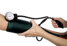
Elderly and
hypertensive patients can experience a significant blood pressure reduction
during and immediately after RT which can lead to fatigue. The blood pressure
fall is probably multifactorial and may be partially attributed to dehydration
and weight loss.
Fatigue can occur as the body repairs the damage to healthy cells and tissues. Some treatment side effects - such as anemia, pain, lack of sleep (insomnia), and rest, and changes in mood - also may cause fatigue.
Rest, energy conservation, adjustment in the dosage of anti-hypertensive drugs, and correcting the above contributing
factors may ameliorate the fatigue. The
following strategies can reduce fatigue and improve the quality of life:
Assess
and document the level of fatigue daily by using a diary or worksheet to
monitor fatigue daily. The fatigue level assessment includes monitoring its
severity (none, minor, moderate, advanced) over the times the day.
Perform
regular daily tasks and activities especially during the time of day when feeling
less fatigue. (based upon one’s diary or worksheet)
Drink plenty of fluids and eat as nutritious as possible.
Avoid caffeine which dries the mouth and can disrupt sleep.
Maintain
a daily exercise program.
Allow plenty of time for sleep each night.
Consult
a social worker or a psychologist, and seek support from family and friends.
Seek
evaluation and treatment of underlying medical and psychological conditions
(i.e., anemia, depression, hypothyroidism).
Try to maintain a positive outlook.
Effect of radiation on the brain
Most RT of head and neck cancer do not
involve the brain. Current radiation technologies spare or minimize the
exposure of the brain to radiation. RT directed at the brain itself
may cause brain swelling.
Short-term side effects include: headaches
(not relieved by acetaminophen), hair loss, nausea, vomiting, poor appetite, extreme
tiredness (fatigue), sleepiness, hearing loss, skin and scalp changes, changes
in vision (i.e., double vision), change in mental status, trouble with memory
and speech, unsteadiness, and seizures.
Treatment
include steroids (to reduce brain swelling) and anti-seizure medications.
Other side effects
Other side effects include trismus and hearing problems (see below) .
Speaking includingtracheoeso-esophageal or esophageal speech, may become more difficult during and immediately
after radiation therapy. This is because of swelling of the vocal cords (in
non-laryngectomees), or the tissues behind the tracheo-esophageal valve; or due
to thick secretions blocking the valve.
Mucous coming up from the stoma is more
sticky and hard to cough up during RT.
LATE side EFFECTS
Late side effects include include permanent loss of saliva; osteoradionecrosis; pharyngoesophageal
stenosis; dental caries; oral cavity necrosis; dysgeusia (taste disorder), fibrosis; impaired wound
healing; skin changes and skin cancer; lymphedema; hypothyroidism; lightheadedness,
dizziness and headaches; secondary cancer; and eye, ear, neurological and neck
structures damage. Patients who undergo radiotherapy for
nasopharyngeal carcinoma tend to suffer from chronic sinusitis.
Permanent dry mouth (xerostomia)
Although the dry mouth (xerostomia) and thick saliva improves in most people with time, it can be long lasting and affects one's quality of life. Xerostomia can lead to the development of
salivary stones and cysts. Permanent xerostomia usually develops a year
after completion of RT is caused by fibrosis of the salivary glands that
follows an initial inflammatory response. Saliva has important function that can be adversely affected following RT. These include: • Lubrication and moistening of food for swallowing • Solubilizing material so it can be tasted • Initiating digestion • Preventing dental caries • Maintaining oral and upper gastrointestinal pH • Supports the health of oral mucosa • Preventing opportunistic infections by maintaining oral microfloral balance • Facilitates speech production • Enhancing denture / prosthesis comfort and function • Cleansing of mouth and clearing the esophagus RT can lead to irreversible salivary glands cell damage. Serous salivary glands (Parotid & submandibular) are very sensitive to radiation. RT often leads to marked changes in the quantity and quality of saliva after just a few doses of radiation and alters the consistency of the saliva from watery to more viscous. Xerostomia can lead to: • Opportunistic infections (mostly fungal such as thrush) • Denture stomatitis • Alterations in pH • Alteration in secretory IgA • Radiation caries (subgingival) leading to tooth decay, gum disease, and oral
infection • Salivary stones and cysts • Burning mouth syndrome (see next section)
Aside from being bothersome to patients, including making it difficult
to eat, swallowing and speak, there is greater risk of dental cavities and dental disease. The maintenance of dentures can become problematic. Patients
who experience low function of their salivary gland and xerostomia must
maintain excellent oral hygiene to minimize the risk of oral lesions.
Periodontal disease can be accelerated and caries can become rampant unless
preventive measures are instituted. Multiple preventive strategies should be
considered. Management and prevention include:
Palliative use of salivary substitutes (gels; rinses)
Dental hygiene (clean mouth multiple times/day and after each
meal, brush teeth, floss, use fluoride toothpaste, rinse with salt/baking soda solution, avoid food with
sugar)
Dentures should be remove and cleaned at least nightly or
as directed by dentist
These measures can help in coping with xerostomia:
Drinking
adequate fluids (increase fluid intake may lead to increase urination), frequent
sipping or spraying of the mouth with water; sucking on ice chips and /or
sugar-free popsicles; consuming acidic or bitter substances, using sugarless
gum and sugarless hard candy can help stimulate saliva, and rinsing and
gargling with diet ginger ale or a weak solution of salt and baking soda are
helpful to refresh the mouth, loosen thick oral secretions, and alleviate mild
pain.
The use of saliva substitutesor artificial saliva (containing hyetellose, hyprolose, xylitol, or
carmellose), or stimulation of saliva production from intact salivary glandular
tissues by taste/mastication, pharmacological sialagogues (a drug that increases the flow rate of saliva),
acupuncture, avoiding smoking and all products that contain caffeine or
alcohol, using a bedside humidifier at night, and raising the head of the bed
can be helpful. Low-level laser treatment can intensify cell metabolism and its application on salivary glands could improve salivation.
Available pharmacologicalmedications include salivary stimulants (sialagogues), such aspilocarpine, amifostine, cevimeline and herbal medications. Pilocarpine is the only drug approved by the U.S. Food and Drug Administration for use as a sialogogue for radiation xerostomia.Preliminary
data suggest that hyperbaric oxygen, hyperthermic humidification, photobiomodulation
/ low-level laser therapy and transcutaneous electrical nerve stimulation can
provides benefit for patients with xerostomia who have some residual salivary
gland function.
Acupuncturecan be used for a dry mouth in people having radiotherapy for head and neck cancers. It is also used for pain and dry mouth after
neck surgery or radiation and a dry mouth in those who had radiation therapy,
and those with advanced cancer. . Several clinical trials suggest that acupuncture can help.
Dietary change from dry, tough food to
easier to swallow moist, softer (i.e., thick soups, mashed potatoes, puddings,
and milkshakes), one can greatly improve nutritional status and quality of
life. Use of a humidification especially in the bedroom can also provide some
relief.
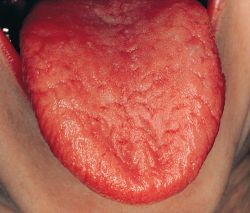
Dry mouth after radiation
Burning mouth syndrome
Chronic or recurrent burning in the mouth
caused by the radiation and enhanced by xerostomia is termed secondary “Burning
mouth syndrome”. Symptoms may include:
A superficial burning or scalded painful sensation affecting
the tongue, lips, gums, palate, throat or whole mouth
Mouth dryness and increased thirst
Loss of taste or changes in taste (e.g.,
bitter or metallic)
The mouth discomfort can come and go or occur
every day. It can be present throughout the day or slowly worsen as the day
progresses. It may last for months to years. Symptoms may suddenly disappear on
rare occasions or become less frequent. Eating or drinking can bring temporarily
relief.
The discomfort can lead to depression, anxiety,
and difficulties in falling asleep and eating.
The discomfort caused by the syndrome can
be reduced by avoiding acidic foods, spicy foods and carbonated beverages, tobacco,
and excessive stress..
Treatment is symptomatic and may include one
or a combination of these methods:
Specific oral rinses or lidocaine
Saliva replacement products
Capsaicin, a pain reliever derived from
chili peppers
Clonazepam or Klonopin, (anticonvulsant medication)
The
risk of dental caries increases after RT of head and neck cancer because for a
number of factors. These include xerostomia that leads to an increase in the number of caries producing bacteria
(Streptococcus mutans and Lactobacillus species) in the mouth,
reduced concentrations of salivary antimicrobial proteins, and loss of saliva's mineralizing components leading to dental deminerilization.
Management strategies includes preoperative
dental treatment, prevention through daily oral care, including fluoride
treatment, and routine visits with dentist/oral hygienist. Optimal oral
hygiene must be maintained and xerostomia should be managed whenever possible by
using salivary substitutes or replacements. Caries resistance can be enhanced
with the use of topical fluorides and/or re-mineralizing agents (high in calcium phosphate and fluoride). The efficacy of topical products may be enhanced by increasing their contact time with the
teeth by using dental trays. Those unable to effectively comply
with use of fluoride trays can be instructed to use brush-on gels and
rinses.
Topical
fluorides or chlorhexidine rinses may lead to reduced levels of S. mutans but not Lactobacillus. Because
of adverse drug interactions, fluoride and chlorhexidine dosing should be
separated by several hours.
Routine oral
care includes: gentle regular brushing using soft bristle toothbrush, use rechargeable
electric tooth brush when possible, brush after eating, rinse with water when unable
to brush, floss, use dental water jet, use fluoride tooth paste, and get
routine dental care prescribed by a dental professional such as fluoride tray.
Osteoradionecrosisof the jaw
This is one potentially severe complication that can necessitate surgical intervention and reconstruction. It is a severe iatrogenic disease of devitalized
bone caused by RT of head and neck cancer and can occur during or after
treatment. It is a state of injured bone tissue with inadequate healing or
remodeling response of at least three to six months. Bone loss or death is the
result of damaged blood vessels within the bone. It can cause bone fracture and
infection.
Depending on the location and extent of the lesion, symptoms may include pain, bad breath, taste distortion (dysgeusia), “bad sensation”, numbness (anesthesia), trismus, difficulty with mastication and speech, fistula formation, pathological fractures, and local, spreading, or systemic infection.Patients who have received high-dose radiation to the head and neck are at lifelong risk for osteoradionecrosis, with an overall risk of approximately 15%.
The jaw bone (mandible) is the most frequently affected bone, especially in those treated for nasopharyngeal cancer. Maxillary involvement is rare because of the collateral blood circulation it receives.
Tooth extraction and dental disease in irradiated areas are major factors in the development of osteoradionecrosis. In some cases it is necessary to remove teeth before RT if they will be in the area receiving radiation and are too decayed to preserve by filling or root canal. An unhealthy tooth can serve as a source of infection to the jaw bone that can be particularly difficult to treat after radiation.
Repair of nonrestorable and diseased teeth prior to RT may reduce the risk of this complication. Oral
disease should be eliminated pretreatment whenever possible. Dentition that exhibits poor
prognosis and is within high-dose radiation fields should be extracted before RT
begins. Ideally, at least 7 to 14 days should be allowed for healing before
initiation of RT; some have suggested allowing up to 21 days.
Mild osteoradionecrosis can be conservatively treated with debridement, antibiotics, and occasionally ultrasound. Topical
antibiotics (e.g., tetracycline) or antiseptics (e.g., chlorhexidine) may
contribute to wound resolution. Pain management may be needed. Wherever possible, exposed bone should be covered
with mucosa and necrotic bone removed. Analgesics for pain control are often effective.When necrosis is extensive, radical resection, followed by microvascular reconstruction is often used.
The combination of pentoxifylline and
tocopherol was effective in treating bisphosphonate and radiation related
osteonecrosis of the jaw. The prophylactic use of the combination reduced
the incidence of osteoradionecrosis following dental extraction.
Hyperbaric oxygen therapy (HBO) has been often used in patients at risk or those who develop osteoradionecrosis of the jaw. However, the available data are conflicting about the clinical benefit of HBO for prevention and therapy of osteoradionecrosis.
HBO
has been reported to increase oxygenation of irradiated tissue, promote
angiogenesis, and enhance osteoblast repopulation and fibroblast function. HBO
is usually prescribed as 20 to 30 dives at 100% oxygen and 2 to 2.5 atmospheres
of pressure. If surgery is needed, ten dives of postsurgical HBO are
recommended. Unfortunately, HBO technology is not always accessible to patients
who might otherwise benefit because of lack of available units and the high
price of care.
Patients should inform their dentists about their RT prior to extraction or dental surgery. Osteonecrosis may be prevented by administration of a series of HBO therapy before and after these procedures. This is recommended if the involved tooth is in an area that has been exposed to a high dose of radiation. Consulting the radiation oncologist who delivered the radiation treatment can be helpful in determining the extent of prior exposure.
Prevention
include adequate dental care and prophylaxis, good nutrition, avoiding alcohol
and tobacco, avoid traumato sift and hard tissues.
Dental prophylaxis can reduce the risk of osteoradionecrosis. Special fluoride treatments may help with dental problems along with brushing, flossing, and regular cleaning by a dental hygienist.
A home care dental lifelong routine is recommended:
1. Flossing each tooth and brushing with toothpaste after each meal.
2. Brushing the tongue with a tongue brush or a soft bristled toothbrush once a day.
3. Rinsing with a baking soda rinse daily. Baking soda helps neutralize the mouth. One teaspoon added to 12 oz. of water. The baking soda rinse can be used throughout the day.
4. Using fluoride in fluoride carriers once a day. Fluoride carriers are custom made by a professional dentist. A 1.1% sodium fluoride or 0.4% stannous fluoride is placed in the fluoride carriers and applied over the teeth for 10 minutes. One should not rinse, drink, or eat for 30 minutes after fluoride application.
Necrosis in the oral cavity
Tissue necrosis
(death of cells) and secondary infection of previously irradiated tissue is a
serious complication for patients who have undergone RT for head and neck
cancer. Acute damage typically involve the mucosa of the mouth.
Chronic changes involving bone and mucosa are a result of the process of
vascular inflammation and scarring that in turn results in tissue damage because
of reduced blood and oxygen supply. Infection secondary to tissue injury and
osteonecrosis confounds the process.
Soft
tissue necrosis can occur in any mucosal surface in the mouth. Trauma and
injury are often associated with nonhealing soft tissue necrotic lesions,
though spontaneous lesions can also happen. Soft tissue necrosis begins as an
ulcerative break in the mucosal surface and can spread in diameter and depth.
Pain will generally become more prominent as soft tissue necrosis becomes
worse. Secondary infection can also take place.
Excessive
depletion may prevent healing and lead to a chronic open wound recognized as soft-tissue necrosis. This may be referred to as a consequential late
effect. Other consequential late effects include mucosal scarring (healing by
secondary intention) and loss of mucosal compliance, contributing to chronic
dysphagia.
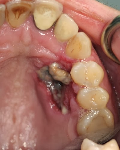
Upper palate necrosis
Fibrosis
High doses of radiation to the head and neck can result in fibrosis where the neck may develop a woody texture and have limited movement. This condition may be aggravated after head and neck surgery. Radiation-induced fibrosiscan develop as a late
effect of RT in area that has been irradiated. RT effects theskin and
subcutaneous tissue, muscles,tendons, nerves, lymphatic system, bones, and
other organs,depending upon the treatment
site. It generally starts eight to twelve weeks after
the initiation of RT and is a lifelong issue. Radiation to the neck can cause significant atrophy and tightness to muscles of the neck
and shoulders including the scalenes, trapezius, and sternocleidomastoid
muscles. Radiation-induced fibrosis may cause both cosmetic and functional
impairment, which can lead to deterioration in the quality of life. Early
intervention to manage fibrosis is very important.
Late onset of fibrosis can also occur in the pharynx and esophagus, leading to stricture, dysphagia and temporomandibular joint problems including mandibular dysfunction. Patients
can be instructed in physical therapy interventions such as mandibular
stretching exercises and the use of prosthetic aids designed to reduce the
severity of fibrosis. It is important that these approaches be instituted
before trismus develops. If clinically significant changes develop, several
approaches can be considered, including stabilization of occlusion, and use of
trigger-point injection and other pain management strategies, muscle relaxants,
and tricyclic medications.
Patients should maintain flexibility of the neck
muscles by stretching exercises including chin curls, head rotations, shoulder
shrugs, and shoulder circles. Exercise can reduce neck tightness and increases
the range of neck motion. One needs to perform these exercises throughout life
to maintain good neck mobility. After radiation fibrosis has developed
individuals may benefits from myofascial release (MFR) if medically
appropriate. MFR is a hands-on method of massaging and stretching the
connective tissue of the head/neck to increase range of motion, increase
flexibility, decrease pain, and improve posture. MFR is typically performed by
a trained speech pathologist or physical therapist. Receiving treatment by
experienced physical therapies who can also break down the fibrosis is very
helpful. The earlier the intervention, the better it is for the patient. There
are physical therapy experts in most communities who specialize in reducing
swelling.
A prospective study ( https://pubmed.ncbi.nlm.nih.gov/30776452/
) showed that pravastatin (a statin) is
an efficient antifibrotic agent in patients with grade ≥2 cutaneous and
subcutaneous fibrosis after radiation therapy for head and neck cancer.
Muscle tightness can often serve as trigger of headaches which may eventually lead to migraine. The muscles of mastication are also often involved.Treatment of muscle fibrosis can often alleviate and reduce the frequency of such headaches.
Neck exercises
A new treatment modality that reduces lymphedema, fibrosis and neck muscle stiffness usingexternal laseris also available. This method uses a low energy laser beam administered by an experienced physical therapist. The laser beam penetrates the tissues where it is absorbed by cells and changes their metabolic processes. The beam is generated by the LTU-904 Portable Laser Therapeutic Unit. Fibrosis in the head and neck can
become even more extensive in those who have had surgery or further radiation. Post radiation fibrosis can also involve the skin and subcutanous tissues, causing discomfort and lymphedema.
Trismus and dysphagia
Fibrosis of the muscles of mastication can lead
to restriction in the range of motion of the jaw with limited mouth opening which
can progress over time. Trismus or
lockjaw (limited opening of the jaw) is common following radiation, especially following
radiation targeting the base of tongue; tonsil; retromolar trigone; soft
palate; temporalis, masseter, and pterygoid muscles; and the temporomandibular joint
(TMG). The prevalence of trismus increases with increasing doses of radiation,
and levels in excess of 60 Gy are more likely to cause trismus. Fibrosis of the
muscles of mastication can lead to the inability to open the mouth (trismus or
lockjaw) which can progress over time.
The normal moth
opening is 35- 45 mm. The severity of trismus varies between mild (30-34 mm
mouth opening), moderate ( 15-29 mm), severe ( 5-14 mm) and profound (0-4 mm).
Trismus can
adversely affect chewing, nutrition, oral and dental care, speech production, and
intubation for future surgery. Trismus impedes proper oral care and treatment
and may cause speech/swallowing deficits. Early massage and prophylactic trismus prevention exercises can be initiated in those considered
at high risk for developing trismus during radiation. These include massage/manual
therapy, passive stretching devices, jaw exercises, and pain management. Treatment
is contraindicated with osteoradionecrosis of the jaw or mandible.
Gentle Jaw stretching exercises,
such as opening the mouth wide like a big yawn and holding 10-15 seconds, is
often the first exercise. If a more aggressive intervention is needed, a speech
pathologist may recommend tongue blade therapy or a device (e.g., Therabite ,
OraStretch, Dynasplint).These devices are increasingly used during RT as a
prophylactic measure to prevent trismus. One of the benefits of these devises
is that they not only stretches the connective tissue that causes trismus, but
also allows for proper mobilization of the temporomandibular joint, thus
addressing a secondary cause of pain and tightness.
The Terabite system
Early treatment of trismus has the potential to prevent or minimize many of the consequences of this condition. As restriction becomes more severe and likely irreversible, the need for treatment becomes more urgent.
A wide array of appliances are available for the treatment of trismus. Devices range widely in cost. Many devices must be custom made for each patient, thus increasing the cost of treatment. Others, such as continuous passive motion devices are rented on a daily or weekly basis. These devices include the following:
Cages that fit over the head
Heavy springs that fit between the teeth
Screws that are placed between the central incisors
Hydraulic bulbs placed between the teeth
The most commonly used treatment is the use of tongue depressors. These are stacked, forced and held between the teeth in an attempt to push the mouth open over time.
Coronoidectomy can be effective at improving trismus refractory to physical therapy in head and neck cancer patients.
The use of palatal
augmentation prosthesis allows reshaping of the hard and/or soft palate to
improve tongue/palate contact during speech and swallowing. This could be a
removable partial denture or complete denture prosthesis.
Swallowing
difficulties due to fibrosis often requires a change in diet, exercise therapy that
include pharyngeal strengthening, range of movement exercises, and isometric
training, and swallow retraining, especially in those who have had surgery
and/or chemotherapy.
Physiological stimulation
include hyperbaric oxygen therapy, Transcutaneous electrical nerve stimulation,
electrotherapy, and biofeedback.
Swallowing exercises are increasingly used as a preventing measure. Partial or total oropharyngeal stricture can occur in severe cases.
Swallowing exercises
The dropped head syndrome
The dropped head syndrome (DHS) is a
disabling condition caused by severe weakness of the neck extensor muscles
causing progressive reducible kyphosis of the cervical spine and the inability
to hold the head up. Dropped head syndrome induced by radiotherapy is very rare
and can occur from 3 months to 30 years after radiation therapy. The mechanism
of late-onset radiation-induced DHS remains unclear. It is thought to result
from primary muscle damage or anterior horn or root lesions at the upper
cervical level within the radiation field.
Treatment with physiotherapy and surgery
have not been very successful, and the management of DHS is supportive,
including employing a cervical collar to maintain the head in an upright
position.The condition generally does not spread or become worse.
Pharyngoesophageal stenosis
Pharyngoesophageal stenosis can be a delayed complication of RT. Pharyngoesophageal (PE) stenosis is an area of narrowing in the pharynx or esophagus. This stenosis can make it difficult to eat (dysphagia), particularly solid food. It can also cause accumulation of saliva and oral secretion in the mouth. If the PE segment becomes completely closed off, the patient will not be able to eat or drink anything by mouth and will need a feeding tube placed directly into the stomach (gastric tube). Treatment of this complication might include frequent placement of dilating catheters down the throat to stretch and open the narrowed segment or by surgically removal of the blocked segment followed by flap reconstruction.
Swallow test radiographs of a high pharyngoesophageal stricture after laryngopharyngectomy
Impaired wound healing
Some patients may manifest wound healing impairment following surgery, especially in areas that have received RT. Some may develop a fistula ( an abnormal connection between the inside of the throat and the skin). Wounds that heal at a slower pace can be treated with antibiotics and dressing changes by specialists.
Skin changes and skin cancer
Patients
treated with RT can experience radiation recall dermatitis. Its estimated frequency is in 9% of
individuals. Patients who suffered from an initial severe dermatitis, may
experience inflammatory waves that can occur weeks to years after their RT. Symptoms of radiation recall are induced by
inflammation in a region that was previously treated with radiation.
The reaction is
characterized by a skin rash typified by redness, swelling, and/or blistering
of the skin. The rash is often painful and can resemble a severe sunburn.
Late-stage or chronic radiation
dermatitis typically presents months to years after radiation exposure. It is
characterized by skin fibrosis, slight color changes to the skin or mild swelling,
atrophy, and widened blood vessels on the skin (telangiectasias).
Individuals
generally lose hair in the region that received radiation (see above).
Radiation
can increase the risk for skin cancers in the area that received radiation. The
most common types of skin cancers seen are basal cell carcinoma and squamous
cell carcinoma. Cancer can also develop in skin flap used for reconstruction
of the hypopharynx. Therefore, it is very important to see a dermatologist and an otolaryngologist regularly. When noticing any changes in the texture or color of the skin in the
radiated area or any new lesions in the field, one should bring those to the
attention of their health care providers for further evaluation.
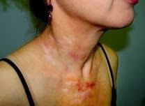
Permanent skin damage after radiation
Radiation recall myositis
Radiation myositis (muscle inflammation) is a rare and infrequent
adverse effect of radiation therapy. It is characterized by muscle tiredness,
weakness and pain, and elevations in muscle enzymes by blood tests (CPK or
aldolase). It can be also diagnosed using MRI, and muscle biopsy.
Skeletal muscle has been considered relatively resistant to
radiation injury. Larger total dose and larger dose per fraction are
influential in the complication rates for muscle injury. Muscle morbidity is
higher for those who received total radiation doses greater than 63 Gy, and may
also be higher in those who receive cytotoxic chemotherapy.
Muscle swelling (edema) following radiation therapy with neutrons
peaks at about six months compared to 12-18 months after treatment with
photons. Edema persists loner in
neutron-treated patients. Complete resolution of photon-induced myositis tends
to occur in about half of the patients within two to three years whereas less than
20% of the patients treated with neutrons have resolution by three to four
years.
Non-steroidal anti-inflammatory drugs are a reasonable initial
intervention for active radiation myositis. The value of corticosteroids for
radiation myositis is anecdotal and controversial. In the presence of severe
tissue breakdown, hyperbaric oxygen therapy may be considered. Hyperbaric
oxygen therapy is of benefit in the treatment of extensive muscle tissue
breakdown due to various causes including radiation injury.
Lymphedema
Obstruction of the cutaneous lymphatics results inlymphedema.Significant pharyngeal or laryngeal edema may interfere with breathing and may require temporary or long term tracheostomy. Lymphedema, strictures, and other dysfunctions predispose patients to aspiration and the need for a feeding tube. Read more about lymphedema at the "Lymphedema, neck swelling, pain and numbness after radiation and surgery" page.
Hypothyroidism
RT is almost always associated with hypothyroidism. The incidence varies; it is dose-dependent and increases as time elapsed since the RT. Read more about it in the Low thyroid hormone (hypothyroidism) and its treatment section.
Hyperparathyroidism
The parathyroid glands are resistant to
radiation therapy. However, hyperparathyroidism (HPT) due to adenoma
formation can occurs in individuals who had received RT for head and
neck cancer after a longer latency period. One third of the patients with HPT
have normal serum calcium levels despite elevated parathyroid hormone levels and
abnormal parathyroid glands.
Signs and symptoms of HPT include:
Fragile bones that easily fracture
(osteoporosis)
Kidney stones
Excessive urination
Abdominal pain
Tiring easily or weakness
Depression and forgetfulness
Bone and joint pain
Frequent complaints of illness with no
apparent cause
Nausea, vomiting or loss of appetite
HPT is diagnosed by finding elevated
calcium levels in the blood, Bone mineral density test (bone densitometry), a
24-hour collection of urine, and imaging tests of kidneys.
Treatment includes watchful waiting in
those with normal calcium levels and kidney functions, and normal bone density.
Medications to treat HPT include calcimimetics (drugs that mimics calcium), hormone
replacement therapy to retain calcium, and bisphosphonates. Surgical removal of the parathyroid adenoma(s)
is curative in most patients.
Parathyroid gland
Attention, thinking, and memory problems (cognitive impairment)
Many patients who received RT to the head and neck and/or chemotherapy experience attention, thinking, or short-term memory problems. Neurocognitive function, although not immediately affected after treatment, progressively declines in 38% of the patients in the 2 years after definitive treatment with chemotherapy or radiation. Other causes for cognitive problems are pain, side effects of medications, emotional state, and other medical problems.
Cognitive problems can manifest in the following symptoms or behavioral changes:
Trouble concentrating, focusing, or paying attention
Mental fog or disorientation
Difficulty with spatial orientation
Memory loss or difficulty remembering things, especially names, dates, or phone numbers
Problems with understanding
Difficulties with judgment and reasoning
Impaired ability to calculation and organize, and impaired language skills. These include difficulties to organize one's thoughts, find the right word, or balance a checkbook
Problems in multitasking
Processing information slower
Behavioral and emotional changes, such as irrational behavior, mood swings, inappropriate anger or crying, and socially inappropriate behavior
Severe confusion
Management of these cognitive problems includes:
Medications, including stimulants, cognition-enhancing drugs, antidepressants, and drugs that block the actions of narcotics
Occupational therapy and vocational rehabilitation, to help people with the activities of daily living and job-related skills
Cognitive rehabilitation and cognitive training, to help patients improve their cognitive skills and find ways to cope with these issues.
Strategies for coping with cognitive problems include:
Keeping a checklist of daily reminders
Doing one task at a time without distractions
Carrying around a small pad and a pen or pencil to write down notes and reminders. Or, downloading a note-making application on one's smartphone and tablet
Using a calendar and a notebook with questions and a to-do list
Letting friends, family, workplace, and health-care team know about one’s memory loss
Getting counseling and other resources to improve memory
Placing sticky notes around the house and workplace as a reminder of important tasks.
Use word play, such as rhyming, to help remember things
Getting plenty of rest
Keeping physically activity to increase mental alertness
Preparing for the next day by setting out the things you will need on the night before
Color coding or labeling certain cabinets or drawers where one stores things around their home
Eliminating clutter, and placing things back in the same place
Neurological damage
Most RT of head and neck cancer do not
involve the brain. Current radiation technologies spare or minimize the
exposure of the brain to radiation. Radiation therapy directed at the brain
itself may cause brain swelling. Late
side effects only affect a small number of adult patients. These effects show
up usually from 6 months to many years after treatment ends. These delayed
effects can include serious problems such as memory loss, stroke-like symptoms,
brain function, and rarely increased risk of having another tumor in the area.
RT to the neck can also affect the spinal cord, resulting in a self-limited transverse myelitis, known as "Lhermitte's sign". The patient notes an electric shock-like sensation mostly felt with neck bending (flexion). This condition rarely progresses to a true transverse myelitis which is associated with Brown-Séquard syndrome(A loss of sensation and
motor function caused by the lateral cutting of the spinal cord) andtypically resolves within one year.
RT may cause neuropathydue to nerve injury, although it may
take several years for symptoms to appear. RT can also cause peripheral nervous system dysfunction resulting from external compressive fibrosis of soft tissues and reduced blood supply caused by fibrosis. Peripheral neuropathy (see Side effects of chemotherapy for head and neck cancer section) is a disorder
that occurs when the nerves outside the brain and spinal cord, called the
peripheral nervous system, are damaged. Depending on which nerves are affected,
symptoms may include a change in sensation, especially in the hands and feet, (e.g.,
numbness, tingling, or pain); muscle weakness, (i.e., myopathy), paralysis and/or paresis (weakness) of the diaphragms
(cause by phrenic nerve damage), and changes in
organ function (i.e., constipation, dizziness).
Radiotherapy-related cranial nerve palsy may
occur after receiving conventional radiotherapy. The most commonly affected
nerves are the hypoglossal, vagus and recurrent laryngeal nerves. If the cranial nerves are affected one
can experience difficulty in speaking, chewing or swallowing; numbness,
tingling, or burning in the jaw, throat, or neck; hearing loss or ringing in
the ears (see below), vision loss or other vision symptoms, such as blurred or
double vision (see below) ; and changes in taste and smell.
The management of neuropathy depend on the
specific symptoms and the location of the nerve pain.
These include:
Medications that treat peripheral nerve
pain, including medicines for nausea and vertigo; certain antidepressants, like
selective serotonin or norepinephrine reuptake inhibitors (SSRIs or SNRIs); or
medications to treat nerve pain, including gabapentin or duloxetine
Nerve blocks and transcutaneous electric
nerve stimulation (TENS)
Speech therapy to improve speech,
chewing, and swallowing
Physical therapy to improve strength,
balance, coordination, or mobility
Occupational therapy to maintain motor
skills needed for daily activities
Damage to the peripheral and autonomic nervous system can lead to dizziness when standing up from sitting up from sitting or lying down due to postural hypotension.
RT can cause some
of the symptoms of dysautonomia (see below).
RT of head and neck cancer seemed to have adverse but insignificant effects on the cognitive functions of the patients.
Dysautonomia
Dysautonomia is
an umbrella term referring to a group of several medical conditions that cause
a malfunction of the Autonomic Nervous System (ANS). This is the part of the
nervous system that controls automatic processes in the body, such as heart
rate, blood pressure, digestion, dilation, and constriction of the pupils of
the eye, kidney function, and temperature control. Dysautonomia can be acute
and reversible or non-reversable, chronic and progressive.
Dysautonomias
in adults often are associated with, and may be secondary to, another disease
process or a drug. Common secondary causes include medications, chemotherapy, radiation
treatments, spinal cord or head injury, diabetes or autoimmune autonomic
ganglionopathy.
Oral, head and
neck cancer patients that receive radiation as part of their treatments, can
have varying degrees of scatter radiation to these structures, and years after
treatment develop some of the manifestations of dysautonomia. When the ANS
doesn’t work as it should, it can cause heart and blood pressure problems,
trouble breathing, and loss of bladder control among other symptoms.
The symptoms
experienced by dysautonomia patients vary based on the type of autonomic
dysfunction and where the dysfunction is occurring within the body.
Symptoms of
dysautonomia
People with dysautonomia have trouble regulating some of the ANS
systems, which can result in lightheadedness, fainting, unstable blood
pressure, abnormal heart rates, malnutrition, and in severe cases, death.
● Balance
Problems
● Chest
Pain/Discomfort
● Fatigue
● Nausea,
Vomiting, GI Trouble
● Irregular
Heart Rate and Blood Pressure
● Fainting/
Loss of Consciousness
● Migraines
● Erectile
Dysfunction
● Noise and
Light Sensitivity
●
Lightheadedness, Dizziness, Vertigo
● Visual
Disturbances
● Weakness
● Sweat less
than normal, or not at all
● Swings in
Body Temperature
● Difficulty
Swallowing
● Brain Fog/
Forgetfulness
● Moodiness
● Sleep
Disturbances
● Frequent
Urination, Incontinence
● Low Blood
Sugar
● Dehydration
● Exercise
intolerance
Patients with
autonomic disorders usually require a multi-disciplinary team, as autonomic
disorders can impact almost every organ and system of the body. While each
specialist will have his or her own areas of expertise, it is still important
for each member of the patient’s team to be familiar with the most up-to-date
information on the patient’s autonomic disorder.
There is no
cure for dysautonomia. Secondary forms of dysautonomia may improve with
treatment of the underlying disease. The best that can be done is to deal with
the various manifestations of it and their individual symptoms. Treating
symptomology is not the same as treating the disease and is a coping mechanism
not a cure. Lifestyle changes will be necessary for many of the symptoms to be
reduced or mitigated.
Plexopathy is a disorder affecting a
network of nerves, blood vessels, or lymph vessels. Radiation-Induced brachial
plexopathy is caused by radiation damage to the brachial plexus, a nerve bundle
near the neck and shoulder. (See picture below). The nerves forming the
brachial plexus originate at the spinal cord in the neck and provide sensation
and muscular innervation for the whole hand.
Symptoms may include paresthesia
(tingling, pricking, numbness), dysesthesia (abnormal sense of touch, such as
burning, itching, feeling of an electric current, “pins and needles”, pain),
decreased sensitivity, partial loss of movement (muscle weakness and difficulty
performing simple tasks such as opening jars, or containers, holding objects),
complete paralysis of the arm, muscular atrophy, impaired mobility and partial
dislocation of the shoulder joint.
The damage to the brachial plexus results
from a combination of direct nerve damage from ionizing radiation, the progressive
development of scar tissue (radiation fibrosis) in and around the nerves, and
damage to blood vessels that supply these nerves. Radiation also causes shrinking
and decrease in elasticity of nerve fibers. The extent of damage is associated
with the radiation dose and technique, and the concurrent use of chemotherapy.
Most patients develop symptoms within the
first three years, however, the interval between the last dose of
radiation and the onset of symptoms ranges between six months and 20 years.
Rehabilitation program includes physical and
occupational therapies. Physical therapeutic modalities emphasis pain lessening,
strengthening, conservation of range of motion, and controlling lymphedema. Occupational
therapy evaluates daily activities and offers suitable adaptive tools.
Brachial plexus
Eye damage Cataract: Radiation
can cause cataract usually in the posterior sub-capsular and sometimes in the cortical
areas of the lens. Several other confounding risk
factors contribute to lens opacities including age, diabetes mellitus,
corticosteroids use, smoking, and ultraviolet radiation exposure. The exact mechanisms of how radiation causes cataract are still not fully understood.
A
number of studies indicate that there is risk of lens opacities at doses below
1 Gy and the threshold may range from none to 0.8 Gy. However, the
International Commission on Radiological Protection (ICRP) has recently
accepted the threshold of 0.5 Gy.
It may take many years or decades before
radiation-induced eye lens injuries become apparent. At relatively high exposures of few Gy, lens opacities may occur within
a few years; however, at lower doses and dose rates (less than 1 Gy), lens opacities
may occur after many years. The duration of the latency period is inversely
dependent on dose.
The
only way to treat cataract is by surgery. This involves removing the opacified
lens, leaving the capsule that contains it intact. A plastic lens is inserted
and, therefore, there is no need to wear special glasses after the operation.
Surgery is only indicated when lens opacity progresses to a stage that causes visual disability.
Radiation retinopathy: This complication can occur following exposure to any source of radiation. It
is especially common after radiation treatment for nasopharyngeal, paranasal
sinus or orbital tumors. Higher total radiation dose has been shown to increase
the risk of radiation retinopathy. The incidence of retinopathy increases at
doses above 45 Gy.
Comorbidities
such as diabetes, hypertension, simultaneous chemotherapy and pregnancy are
associated with an increased risk of radiation retinopathy.
Appropriate
shielding of the ocular structures during external beam radiation and hyperfractionation
of external beam can decreased the incidence of radiation retinopathy.
Patients
with early or mild retinopathy may be asymptomatic. Those with advanced disease can present with decreased vision or floaters.
Treatment
includes intravitreal injection of humanized monoclonal antibody to vascular
endothelial growth factor (Bevacizumab), intravitreal triamcinolone acetonide,
grid macular laser photocoagulation, sector scatter and pan-retinal laser
photocoagulation, photodynamic therapy, hyperbaric oxygen and oral
pentoxifylline. Advanced proliferative radiation retinopathy complicated by
vitreous hemorrhage, and/or tractional retinal detachment may require vitrectomy.
Radiation neuropathy
Damage to the ear (ototoxicity) and hearing loss
Patients
who undergo radiation therapy for head and neck cancer appear more likely to
experience hearing loss and to be more disabled by its effects than those who
do not receive such treatment. Common complaints are ear heaviness, earache, decreased hearing, tinnitus, and dizziness.Dose of radiation is directly proportional to ototoxicity; minimum 60 Gys of total
radiation dose is required to produce significant ototoxicity.
Chemoradiation therapy can cause progressive hearing impairment especially in those receiving the chemotherapy intravenously, with a modest deterioration of 5 decibel 4.5 years after treatment.
Radiation to the ear may result
in serous otitis (otitis with effusion). This condition is associated with accumulation of fluid in the middle ear and temporary reduced hearing. Serous otitis and conductive deafness arereversible over time. High doses of radiation can cause sensorineural hearing loss due to damage to the
inner ear, the auditory nerve, or the vestibular apparatus.This condition is not reversible.Damage to the vestibular apparatus can cause dizziness and vertigo.
Hearing loss may make individuals perceive that their
voice is weaker than it actually is. It may contribute to social isolation and
make laryngectomees try to speak louder. In those who use tracheoesophageal
speech and are trying to speak louder by generating greater expiratory effort a
separation of the HME base plate from the skin can occur. This is especially true in those who use a hands
free HME. Wearing a hearing aid
enables a laryngectomee to hear their actual voice.
Radiation-induced chronic rhinosinusitis RT-induced toxicity to sinonasal mucosa can lead to impaired nasociliary
clearance as well as impaired mucous production (xerorhinia).This can lead to production of thick
secretions with decreased nasal clearance. Individuals experience thick,
foul-smelling crusts that block their nasal passages. Management of this chronic condition often requires frequent debridement, nasal irrigation, and a
nasal steroid.There is a high prevalence of recovery of methicillin
resistant Staphylococcus aureus (MRSA) from these sinuses. It is
advisable that appropriate cultures are obtained from patients so that proper
antimicrobials can be administered.
Lightheadedness, dizziness and headaches
Lightheadedness,
dizziness, and headaches can be one of a late side effects of radiation of the
head and neck. Damage to the peripheral and autonomic nervous system and the carotid artery baroreceptors can lead to dizziness when standing up from a sitting or lying down, due to the development of low blood pressure (orthostatic hypotension). This can be prevented by standing up slowly, wearing
of compression stockings, exercising and keeping well hydrated. It is best to
consult one’s physician to prevent and treat this condition. Lightheadedness can also be caused by Postprandial Hypotension (see below).
The
perception of the body’s position is determined by the brain by integrating
information from the middle ear, eyes, and the body’s muscles and joints. Unfortunately,
radiation almost always causes head neck muscles fibrosis and can also infrequently
damage the middle ear. The perception of lightheadedness and dizziness after
radiation treatment may be generated in some individuals by misinformation sent
to the cerebellum (the part of the brain that controls the body’s balance) from these locations.
Dizziness
and lightheadedness can be treated by physical therapy. This includes vestibular
rehabilitation and exercises that stretch the fibrotic muscles, reduce neck stiffness,
and increase the head and neck range of motion. One needs to perform these
exercises throughout life to maintain good neck mobility.
Vestibular rehabilitation therapy is an exercise-based program designed to promote
central nervous system compensation for inner ear deficits as well as
misinformation sent to the brain from other parts of the body (eyes, skin, muscle,
joints etc).
Dizziness
and lightheadedness can be caused by a variety of causes and condition and
should be evaluated by one’s physician and medical specialists (e.g., neurologist, otolaryngologist).
Muscle
tightness and fibrosis can often serve as trigger of headaches which may
eventually lead to migraine. The muscles of mastication are also often involved. Treatment of muscle fibrosis can often alleviate and
reduce the frequency of such headaches.
Damage to neck structures
Neck edema and fibrosis are common after RT. Over time the edema may harden, leading to neck stiffness. Damage can also include carotid artery narrowing (stenosis) and stroke, carotid artery rupture, oropharyngo-cutaneous fistula (the last two are associated also with surgery), and carotid artery baroreceptors damage leading to permanent and proxysmal (sudden and recurrent) hypertension. Ischemic stroke
can be a late complication of neck irradiation. Many factors can contribute to
the increased risk of stroke in patients with head and neck cancer, including
carotid artery stenosis (see below) and increased deposition of plaque, as well
as other preexisting risk factors for cerebrovascular disease, particularly
smoking.
Carotid artery stenosis and carotid artery (CA) rupture:The carotid arteries in the neck supply blood to the brain. Radiation to the neck has been linked to CA stenosis or narrowing,and rarely to CA rupture; representing a significant risk for head and neck cancer patients, including laryngectomees. Screening ultrasound within the first year since completion of radiotherapy, followed by repeat ultrasounds every two to three years and whenever CA stenosis is suspected can lead to early diagnosis. Smoking increases the risk of CA stenosis. The incidence of carotidarterystenosis ranged from 18 to 38% in patients who underwent radiotherapy for head and neck cancer compared to 0 to 9.2% among nonirradiated patients.CA disease can cause strokes and transient ischemic attack (TIA), though it does not always cause symptoms.It is important to diagnose carotid stenosis or impending rupture early, before a stroke or severe bleeding has occurred.
internal carotid artery 90% stenosis (left) and stenosis repaired with a stent (right)
Stenosisincreases the
risk of stroke, which is a medical emergency that occurs when the blood supply
to the brain is interrupted or significantly decreased.
The signs and symptoms of a stroke or TIA include these sudden features:
Numbness or weakness in the face or
limbs, often on only one side of the body
Difficulty in speaking and understanding
Trouble seeing in one or both eyes
Dizziness or loss of balance
Severe
headache with no known cause
Stenosis can be diagnosed by hearing a bruit sound over the CA,ultrasound, CT, MRI, MRA (Magnetic Resonance Angiogram ) as well as angiography. Treatment of stenosis caused by RT is usually by placing a stent (a small device placed inside the artery to widen it). Other methods include removal of the blockage (endarterectomy), or a prosthetic carotid bypass grafting.
Evidence suggesting impending rupture can be obtained on physical and radiological examinations. Endovascular stenting is also performed in patients with impending CA rupture.
Hypertension and hypotension due to baroreceptors damage:Radiation to the head and neck can damage the baroreceptors located in the carotid artery. These baroreceptors help in regulating blood pressure by detecting the pressure of blood flowing through them, and sending messages to the central nervous systemto increase or decrease the peripheral vascular resistanceand cardiac output. Some individual treated with radiation develop labile or paroxysmal hypertension.
Low blood pressure (hypotension): This can be cause by damage to the peripheral and autonomic nervous
system and the carotid baroreceptors.This can lead to baroreceptor failure that is manifested in orthostatic hypotension (intolerance) which is characterized by dizziness
when standing up from a sitting or lying down.This can be managed by standing up slowly,
wearing of compression stockings, exercising and by keeping well hydrated. Hypotension can also be caused by by Postparndial Hypotension (see below)
Labile hypertension: In this condition the blood pressure fluctuates far more than usual within the day. It can rapidly soar from low (e.g.,120/80 mm Hg) to high (e.g., 170/105 mm Hg). In many instances these fluctuations are asymptomatic but may be associated with headaches. A relationship between blood pressure elevation and stress or emotional distress is usually present.
Paroxysmal hypertension:Patients exhibit sudden elevation of blood pressure (which can be greater than 200/110 mm Hg) associated with an abrupt onset of distressful physical symptoms, such as headache, chest pain, dizziness, nausea, palpitations, flushing, and sweating. Episodes can last from 10 minutes to several hours and may occur in different frequency - varying from once or twice daily to once every few months. Between episodes, the blood pressure is normal or may be mildly elevated. Patients generally cannot identify obvious psychological factors that cause the paroxysms. Direct massage of the carotid artery during Doppler ultrasound can lead to such episodes. Medical conditions that can also cause such blood pressure swings need to be excluded (e.g. pheochromocytoma).
Both of these conditions are serious and should be treated. Management can be difficult and should be done by experienced specialists. Correction of carotid artery stenosis (i.e., stent
insertion) can prevent or reduce the occurrence of these conditions.Dr Samuel Mann from Weill-Cornell Medical Center in New York City is one of the leading experts in treating paroxysmal hypertension. The Vanderbilt Autonomic Dysfunction Center is
one of the nation's leading expert centers in dysautonomia, or autonomic
dysfunction.
Determining the accurate BP is essential to provide proper management. To find the true BP it is important to
follow these instructions. Unfortunately these guidelines are often not
followed in many physicians’ offices. It is important that patients insist that they
are followed by medical professionals.
Be still. Don't smoke, drink caffeinated beverages or exercise within 30
minutes before measuring your blood pressure. Empty your bladder and ensure at
least 5 minutes of quiet rest before measurements. Laryngectomees using
esophageal speech or trachea esophageal speech should avoid speaking for at
least 5 minutes.
Sit correctly. Sit with your back straight and supported (on a dining chair,
rather than a sofa). Your feet should be flat on the floor and your legs should
not be crossed. Your arm should be supported on a flat surface (such as a
table) with the upper arm at heart level. Make sure the bottom of the cuff is
placed directly above the bend of the elbow. Check your monitor's instructions
for an illustration or have your healthcare provider show you how.
The ideal cuff bladder length is ≥ 80 percent of the patient’s arm circumference.
The ideal cuff bladder width is ≥ 40 percent of the patient’s arm circumference.
Measure at the same time every day. It’s important to take the readings at the same time each day,
such as morning and evening. It is best to take the readings daily, however,
ideally beginning 2 weeks after a change in treatment and during the week
before your next appointment.
Take multiple readings and record the results. Each time you measure, take two or three
readings one minute apart and record the results.
Don't take the measurement over clothes.
Low blood pressure after eating in individuals
with baroreceptor damage after head and neck radiation ( Postprandial
hypotension).
Low blood pressure after eating can occur in
some individuals with baroreceptor damage caused by radiation therapy for head and neck cancer. This can make them feel dizzy, light headed, and cause them to faint when they
get up quickly.
Prevention and ameliorating of the low blood
pressure after eating can be done by:
Drinking liquids before meals. Drinking 12 to 18 ounces of water 15 minutes before eating can
blunt a fall in blood pressure.
Eating smaller meals. Larger meals are more likely to cause postprandial hypotension
than smaller ones. Try changing from three meals a day to six or seven smaller
meals.
Consuming fewer rapidly digested carbohydrates. White bread and other foodstuffs made with refined flour, white
rice, potatoes, and sugary beverages move rapidly from the stomach to the small
intestine. This quick passage causes postprandial hypotension. Reducing the
consumption of these foodstuffs and substituting them with slowly digested
whole grains, beans, protein, and healthy oils may keep the blood pressure adequate
after a meal.
Moving slowly and carefully. Blood pressure usually is the lowest 30 to 60 minutes following a meal. Sitting or lying down for about an hour after eating can help with
postprandial hypotension. When moving around, one has to use caution and be alert
for signs of low blood pressure.
Several medications and supplements have been
tested to treat and prevent postprandial hypotension. These include caffeine,
guar gum (a water-soluble fiber), acarbose (a diabetes drug that slows the
digestion of carbohydrates), midodrine (increases blood pressure), and others. However,
none of those worked well in clinical trials, and side effects of these
therapies may be significant.
When feel unwell after eating, one should consult
their physician who can properly diagnose and treat them.
Secondary
cancers
Even though radiation is used to treat cancer, paradoxically, years
later it can rarely result in new local and systemic cancers appearing. The risk is proportional to the administered radiation
dose. The secondary cancer can be quite
different from the original and could include local cancers such as skin, mediastinal, oral and thyroid cancer, and systemic cancers such as lymphomas, sarcomas and leukemia. It is important to be closely followed by one's physician as well as medical specialists (i.e., dermatologist) to detect secondary malignancies.